Otoplasty for Prominent Ears
Prominent ear, also known as otapostasis or bat ear is an abnormally protruding human ear. In this condition, the concha (the depression in the external ear leading to its central opening) is large with poorly developed antihelix (a curved prominence of cartilage parallel with and in front of the helix on the pinna) and scapha (the curved depression separating the helix and antihelix of the ear).
Approximately 1-2 % of the total population is affected by prominent ears problem. It is an inherited problem and may be unilateral or bilateral and could be a result of lack or malformation of cartilage during primitive ear development. 30% of babies who have prominent ears are born with normal ears. It is only after 3 months that the problem starts to arise and can get worse when the soft cartilage is repeatedly bent over during breastfeeding.
Causes of protrusion of the external ear:
- Underdevelopment or effacement of the antihelix
- Over development of the deep concha, or
- A combination of both of these features
Even though prominent ears don’t have any functioning issues, it could cause psychological problem in the child & can lead to reduced self-esteem & poor performance in school.

Have questions or want to get started? We are ready to help you with a smile!
Treatment – Otoplasty
Otoplasty is a surgical treatment done to set the prominent or protruding ears back closer to the head or to reduce the size of large ears. It can be carried out in the child from the age of 8 years & above. The surgery can also be carried out in adult patients.
In prominent or Protruding ears, the antihelix is not properly formed or is completely missing. To correct this, antihelix has to be made. Some patients have prominent ear of a smaller grade, in which the prominent ear has the antihelix form but that antihelix at the middle portion is not more prominent. So to reconstruct that antihelix by suturing method is performed.
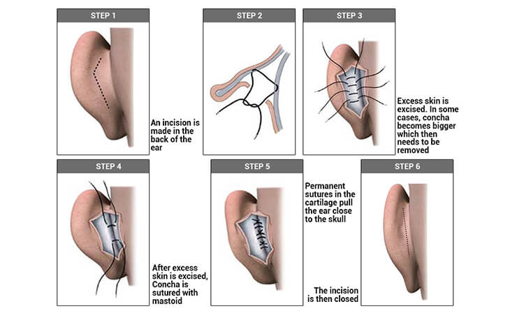
Otoplasty is usually done under a general anesthesia in children & local anesthesia in adults. Once the patient is anesthetized, an incision is made behind the patient’s ear in the fold of the skin where the ear meets the head. Through this incision, extra skin is excised. After this, Concha is sutured with mastoid (bone behind the ear), its called concha mastoid sutures, so the ear sticks towards the back.
Each patient is different for prominent ear. In some patients the inner part of antihelix, the concha becomes bigger which then needs to be removed. In some patients, the angle of the fold in the ear cartilage may cause the ear to protrude, or in some, the ear lobe may be unusually large.
Dr. Rajat Gupta
MBBS, MS, DNB(Gen. Surg.),
DNB (Plastic Surgery)
Dr. Rajat Gupta is a board certified plastic surgeon in India with 10 years of experience to back his expertise in the domain of aesthetic surgeries.
Having completed his training from Maulana Azad Medical College and equipped with a thorough understanding of aesthetic needs of people, Dr. Gupta strives to offer the best remedies and cosmetic procedures outfitted with the latest technology to the aspirants in India and across the globe. To book an appointment, call: +91-9251711711 or email: contact@drrajatgupta.com


