
A Comprehensive Guide to Ear Reconstruction Surgery
There are five basic senses: sight, hearing, smell, taste, touch. Without any of these senses, a person is impeded in today’s world. We live in a highly sensory world. With technology’s constant progress and everything becoming touchscreen, it is no surprise that sensory overload can happen just from watching an intense movie.
But what happens when you lose one of the senses? Say, you lose your sense of hearing? Or it gets impeded in some way. Well, this is common enough. What is less common, however, is the loss or underdevelopment of the external ear.
This is what we want to discuss through this article – the ear reconstruction surgery.
Ear reconstruction is a form of surgery that is performed by expert surgeons to rebuild an ear, which could be damaged by a physical trauma or could be a result of other severe illness like cancer. A missing ear is also sometimes a result of a congenital (present at birth) disorder. With time, this particular surgery has gained importance as it helps many to revive their lives.
To understand the ear reconstruction surgery, let us first expand on the anatomy and importance of an ear.
The Role of the External Ear
While you may have a general understanding about the anatomy of the ear, i.e., the sound waves go through the canal and hit the eardrum, which then sends signals to the brain, you need to know the nitty-gritty to understand why this is such a huge deal.
The external ear plays a central role in conducting sound and directing it towards the eardrum. It essentially helps catch the sound waves so that they come to you, and you hear them.
Additionally, you should also note that ears play a huge role in your balance. The canals are filled with fluid and lined with tiny hairs. When your head moves, the fluid in the canals sloshes around, moving the hairs. The hairs send this position information as signals through the vestibular nerve to your brain. Therefore, you manage to maintain your balance.
The external ear plays the role of an acoustic antenna: the pinna (together with the head) diffracts and focuses sound waves, the concha and the ear canal act as a resonator.
The external ear can be divided functionally and structurally into two parts; the auricle (or pinna) and the external acoustic meatus – which ends at the tympanic membrane (the eardrum).
Have questions or want to get started? We are ready to help you with a smile!
The Auricle
The auricle or pinna directs sound waves towards the external acoustic meatus. It is a mostly cartilaginous structure, with the lobule being the only part not supported by cartilage.
In the middle of the auricle is a hollow depression, called the concha. It continues into the skull as the external acoustic meatus. The concha directs sound into the external acoustic meatus.
The External Acoustic Meatus
The external acoustic meatus is a sigmoid-shaped tube that extends from the deep part of the concha to the tympanic membrane. The walls are 1/3 formed by cartilage, while the inner 2/3 is the temporal bone.
Tympanic Membrane
Finally, the sound reaches the tympanic membrane or the eardrum. The membrane directs it to the middle part of the ear and then to the deepest nerves. These send signals to your brain and help you understand what you just heard.
This way, sound travels. But imagine if it was stopped at the first step itself – at the pinna. If you have no pinna, it is much harder to direct sound. The pinna essentially acts as an amplifier, making it necessary for your brain to begin its comprehension process.
This condition is known as Microtia or Anotia.
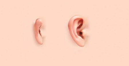
What is Microtia?
Microtia is a congenital deformity (birth defect) in which the external ears are underdeveloped or malformed. Although uncommon, this can cause many problems and unhappiness for the parents, who want to see their children happy and healthy.
Microtia may be seen in one ear or both ears. When in one ear, it is called unilateral, while in both ears, it is bilateral. There are four grades of microtia severity:
- Grade 1: The external ear appears small and mostly healthy, but the ear canal may be narrowed or missing.
- Grade 2: The bottom third of the ear, including the earlobe, appears to be normally developed, but the top two-thirds are small and malformed. The ear canal may be narrow or missing.
- Grade 3: This is the most common type of microtia observed in infants and children. Parts of the external ear may be underdeveloped, including a lobe’s beginnings and a small amount of cartilage at the top. With this grade, there is usually no ear canal.
- Grade 4: The most severe form of microtia is also known as anotia. Your child has anotia if there is no ear or ear canal present, either unilaterally or bilaterally.
While microtia primarily affects children, it can also be present in adults, caused by some trauma. In such cases, ear reconstruction surgery is necessary for the best results.
Ear Reconstruction Surgery
Ear reconstruction surgery is highly sophisticated since it depends on an innate knowledge of how an ear should look. When a surgeon performs reconstructive surgery, they should also ensure natural-looking results.
This is why the procedure is so complicated: it requires the accurate shaping of the ear. The cartilage used to form the external ear needs to resemble an actual ear. Then, when placing it, it needs to be accurately placed so that it looks good and ensures the patient can maintain proper balance.
So, now your next question may be, how is the external ear created? Well, it is created through harvesting cartilage from the ribs and using it to create ears.
Rib Cartilage Ear Surgery
Rib cartilage ear surgery is one of the most commonly used techniques for ear reconstruction. The surgery involves removing a portion of the patient’s rib cartilage and carving it to create a new ear framework. This framework is then placed in a pocket under the scalp skin.
Rib Cartilage ear surgery might need 2-4 surgeries for the optimum results, but highly skilled surgeons can achieve optimum results in the least number of surgeries.
This surgery is age-dependent; the patient needs to be at least eight years old. By this age, most potential patients have developed the necessary rib cartilage for the ideal results. However, if this is not the case, the surgery may be delayed until there is enough disposable cartilage to create a symmetrical ear. Generally, once the chest circumference reaches 60cm, the surgery can be performed.
The Procedure
The procedure involves harvesting the rib cartilage from the patient. This leaves a scar of about 6-8cm in length, but it does not deform the patient’s chest, nor does it hamper sports. It simply allows the surgeon to take the necessary cartilage to reshape then to fit you and your ear.
The new ear is sculpted from the patient’s cartilage and skin. It is placed under the skin to stay alive and continue growing normally with the child, therefore healing without any severe problems. This allows the patient to resume regular sports, swimming, and PE activities 4-6 weeks after surgery with no particular concerns.
The procedure itself has two parts to the operation. Although this may need up to 4 sessions to complete, the necessary 2-step operation process remains the same regardless of how many sessions each operation takes.
First Operation – Getting the Cartilage
During the first operation, the rib cartilage is harvested, and a framework is prepared that is then implanted beneath the skin. The ear framework is specifically done to resemble the other ear as much as possible. In cases where the condition is present in both ears, both frameworks need to match. Meanwhile, if only done in one year, the framework needs to look like the existing, formed ear, for the best results.
In this stage of the surgery, all the ear’s contours will be reproduced, but there will be no space behind the ear. Therefore, the ear appears normal from the side profile but will not stand out like the other.
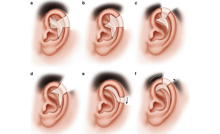
The Second Operation – Making the Results Look Natural
It is in this step that the extra, aesthetic step is taken for the best results. To make the ear look symmetrical to the other ear, a second operation is done to create post-auricular sulcus (space behind the ear) that makes sure that both ears match when viewed head-on.
This is very important – it ensures that you get symmetrical and aesthetic ears. This way, even while the child grows, the years usually grow and continue looking natural.
The interval to be observed between these two operations ranges from three to six months, depending on the particular patient’s situation.
Understanding why cartilage is so important also matters. You need a surgeon who understands your needs and knows the kind of procedure you require. This involves understanding the ideal age for cartilage harvesting so that the ear can be shaped.
Remember, ears serve several purposes, and they need to be symmetrical. If you wear glasses, you will understand this better – uneven ears result in lopsided glasses. Avoiding this is your surgeon’s job. This is why, with the right surgeon, you get all the benefits of rib cartilage surgery.

Benefits of Rib Cartilage Surgery
First and foremost, the created ears are made out of cartilage taken from the patient’s ribs. Therefore, the cartilage is 100% compatible with the body and is alive, growing with the child.
That said, there are also several other benefits to the rib cartilage surgery. Although we have touched upon most of them, let’s go through them once again, shall we?
The Guaranteed Safety
The primary reason rib cartilage surgery is desirable is due to its guaranteed safety. It is entirely safe because there are no artificial substances inserted into the body. In some surgeries, surgeons use implants instead of the rib cartilage technique. This can have adverse results depending on the patient’s immune system and allergies. Such factors determine bodily response to a new substance – the implant – entering the body.
However, with rib cartilage, there is no chance of anything going wrong. Much like fat grafting, which utilises fat from the patient’s body for enhancement, the rib cartilage surgery uses cartilage that has already proven to be compatible with the patient’s body.
Therefore, it is a very safe procedure as the body’s natural tissue is used instead of a manufactured product.
Development of High-Quality Techniques
Understanding the commonplace use of this technique ensures that you also appreciate its quality. Today, rib cartilage surgery has become the standard procedure for Microtia. It is the most reliable and safe method of giving the child the ears they deserve. This means that the techniques surrounding it have also evolved.
For example, surgeons make the smallest incision possible when harvesting the cartilage. This way, they can ensure that no surgical scars are hindering the patient in any way.
Lifelong Results
The best part about using the patient’s rib cartilage is that it is still alive. When the new ear is placed under the skin, it is living. This means it can continue growing like the rest of your body. As the child grows, both their ears increase in size simultaneously – the new one does not stay small.
Therefore, this procedure gives your child lifelong results that never look unnatural.
No Issues with Physical Activity
Generally, after surgery, there is a possibility of physical activity being impeded. This is not something your child experiences after their ear reconstruction surgery. The development of the rib cartilage procedure ensures that it has no significant adverse effects, and they can return to all their physical activity as soon as they want to.
Getting High-Quality Results
All these benefits boil down to one major aspect – the kind of results you get. With a surgeon who understands all these benefits, the chances of you getting the desired, symmetrical results are much higher.
The ideal results can be a source of a lot of joy among families. Seeing their child have both ears in perfect symmetry is often a dream come true. Additionally, the child’s life also is much more comfortable. It is an unfortunate reality that we live in where lacking something makes you “abnormal”. If the child gets these high-quality, natural-looking ears, their life is usually much easier later on.
Of course, these high-quality results are entirely dependent on the kind of surgeon you have. Optimum results can only be achieved when an experienced surgeon performs the procedure. This brings us to the final section of this blog: the standards you need to have to get these kinds of results.
Choosing the Right Centre
With the surgeon comes the centre. You need a well-equipped, high-quality centre for the best results—a centre which ascribes to the highest levels and standards of patient care. Remember, you are going for surgery. You deserve complete care. You should get the kind of attention you deserve. Therefore, your centre matters.
This is also another measure by which to judge your surgeon. If they claim to have all the expertise in the world and then take you to a sub-par centre, then it means nothing. Their claims need to amount to something, and the centre is representative of that.
Choosing the Right Technique
Finally, any procedure concerned with the ear is highly technical. Your ears are some of the most sensitive parts of your body. They need to be handled correctly during surgery; otherwise, more damage can come to them. Therefore, your surgeon’s technique matters. They need to be precise and only cut and reshape as much as necessary.
Only through such extreme care do you get the best results. You should not settle for anything less, simply because ear reconstruction means reconstructing your life to a certain extent.
With Dr Rajat Gupta, at RG Aesthetics, you will not have to! Well-versed in ear reconstruction, he has the delicate and careful fingers necessary to handle such a vital organ. As a leading specialist in ear reconstruction and rib cartilage surgery, he has the required expertise to ensure his patients always get the best, most natural-looking results from their ear reconstruction surgeries under his care!
Dr. Rajat Gupta
MBBS, MS, DNB(Gen. Surg.),
DNB (Plastic Surgery)
Dr. Rajat Gupta is a board certified plastic surgeon in India with 18+ years of experience to back his expertise in the domain of aesthetic surgeries.
Having completed his training from Maulana Azad Medical College and equipped with a thorough understanding of aesthetic needs of people, Dr. Gupta strives to offer the best remedies and cosmetic procedures outfitted with the latest technology to the aspirants in India and across the globe. To book an appointment, call: +91-9251711711 or email: [email protected]
Share this post
Related Posts

What Is Ear Pinning and What Does It Do?
Have you ever been conscious about the way your ears stick out?... read more

Ear Reconstruction: The Innovation That Is Changing Lives!
Ear reconstruction, today, is encompassing all the treatments and surgeries used for... read more
Ear Lobe Repair Surgery: Getting Ear Back In Shape!
Do you avoid wearing big traditional earring due to stretched or torn... read more
Microtia – Causes, Types & Treatment
Microtia is a medical term used for congenital deformity in which a... read more

Can You Hear After Microtia Surgery?
In a world filled with symphonies, whispers of love, and the joyful... read more

Can You Fix the Microtia?
Are you tired of feeling self-conscious about your ear's appearance? Do you... read more